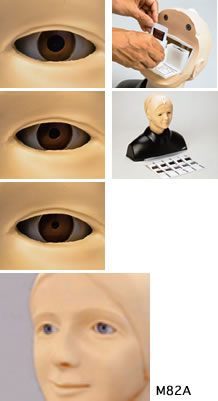
M-82,M82A
;void(0);)
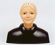
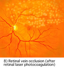
The EYE Examination Simulator is an innovative trainer for fundus examination, designed to allow examination of eyegrounds with the physician's own ophthalmoscope. Various cases can be set up for trainees using combinations of choice of slides, depth and pupil diameter. Soft and supple material allows hands-on simulation of real examination procedures, such as raising the eyelid.
Production Supervision:Japan Society for Medical Education Working Group with the cooperation of: Kansai Medical University Department of Ophthalmology
|
Last ned produktbeskrivelse (PDF)
M82A Se video (WMV) 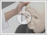
|
Funksjoner
|
|
|
SpecificationsSet includes:
|
|
Manikin size:42 x 21.5 x 38H cm, 2 kg Packing size:30 x 70 x 50H cm, 10 kg (M82A : 35 x 47 x 25 cm, 3.5 kg) *M82A Eye Examination Simulator II is for USA&Canada customers only. *An otoscope is not supplied with the unit. *Specifications are subject to change. |
|
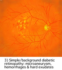
CasesCases:
|
|
Replacement partsReplacement parts:
|
|
Teknisk informasjon
The classification of lung sounds is based on the criteria of the American Thoracic Society.
| 36 cases are available for training. 34 cases include 2 versions -- with and without heart-sounds | |||
|---|---|---|---|
| NORMAL | standard | FINE CRACKES | both lower area |
| mildly weak | both lower and middle area | ||
| mildly strong | whole thorax 1 | ||
| mildly rapid | whole thorax 2 | ||
| loud heart sounds | WHEEZES | upper and middle area | |
| ABNORMAL | weak: left lower area | around trachea and upper area1 | |
| weak: left whole area | around trachea and upper area2 (polyphone) | ||
| absent: left | RHONCHI | trachea and upper area | |
| weak: right lower area | trachea and upper area (polyphonic) | ||
| weak: right lower area | with an inspiratory wheeze | ||
| absent right | whole thorax | ||
| weak: whole thorax | MISCELLANEOUS CONTINUOUS SOUND |
stridor | |
| bronchial sounds | squawk | ||
| COARSE CRACKELS | right lower area | MISCELLANEOUS | pleural friction rub: left lower area |
| both lower area | pleural friction rub: right lower and middle area | ||
| right middle area | Hamman's sign | ||
| left lower area | Vocal fremitus (palpable at both sides of the chest) | ||
| both upper area | |||
| whole thorax | |||
Components & Specifications
| Component | Qty | Measurements | Packing size | Specifications |
|---|---|---|---|---|
| LSAT model unit | 1 | 32 x 35 x 62H cm | 51 x 46 x 80 cm 10 kg | Torso with rotary base 15 built-in speakers 8 ch. amplifier |
| PC | 1 | 59 x 59 x 40 cm 15 kg | Windows XP, 12ch.D/A PCI board,mouse, 112keyboard, 15"TFT monitor *Software & data installed |
|
| Amplifier | 1 | 32 x 35 x 8H cm | 46 x 46 x 15 cm 10 kg | AC 120-240V |
| Speakers | 2 | 62 x 41 x 40 cm 20 kg (incl. monitor) |
||
| T-shirt | 1 | free size |